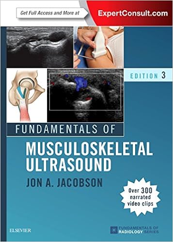
13 best radiology ultrasonography
Radiology ultrasonography, commonly known as ultrasound or sonography, is a medical imaging technique that uses high-frequency sound waves to create real-time images of the inside of the body. It is widely used in the field of medicine and health sciences for diagnostic purposes. Here are some key aspects of radiology ultrasonography:
Principle of Ultrasound: Ultrasound imaging is based on the principle of sending high-frequency sound waves (ultrasound waves) into the body and receiving the echoes that bounce back. These echoes are then processed to create images.
Transducer: A crucial component of ultrasound is the transducer, which is a handheld device that emits and receives ultrasound waves. The transducer is placed on the skin's surface in the area of interest.
Real-Time Imaging: One of the significant advantages of ultrasound is its ability to provide real-time images. As the transducer moves or the patient's body changes position, the ultrasound machine continuously updates the images.
Non-Invasive: Ultrasound is a non-invasive imaging technique, meaning it does not require the use of ionizing radiation (as in X-rays) or invasive procedures like surgery. This makes it safe and widely used for various medical conditions, including during pregnancy.
Applications: Radiology ultrasonography has a wide range of applications in medicine. It is commonly used for imaging the abdomen (e.g., liver, kidneys, gallbladder), pelvis (e.g., reproductive organs), heart (echocardiography), blood vessels (vascular ultrasound), and musculoskeletal system.
Obstetrics and Gynecology: Ultrasound is extensively used in obstetrics to monitor fetal development during pregnancy.It can also detect gynecological conditions and abnormalities.
Guided Procedures: Ultrasound is used to guide medical procedures such as biopsies and needle aspirations. The real-time imaging helps ensure accuracy during these interventions.
Doppler Ultrasound: Doppler ultrasound is a specialized form of ultrasound that assesses blood flow. It is valuable in evaluating vascular conditions, heart function, and identifying blood clots or blockages.
Advancements: Advances in technology have led to 3D and 4D (real-time 3D) ultrasound imaging, providing more detailed and lifelike images. These advancements have improved the diagnosis and assessment of various medical conditions.
Patient Preparation: In many cases, no special preparation is needed for an ultrasound. However, depending on the area being examined, patients may be asked to fast (for abdominal ultrasounds), drink water (for pelvic ultrasounds), or follow specific instructions.
Interpretation: Ultrasound images are interpreted by trained healthcare professionals, such as radiologists or sonographers. They analyze the images to make diagnoses or assess the condition being examined.
In summary, radiology ultrasonography is a valuable medical imaging technique that uses sound waves to create real-time images of the body's internal structures. Its non-invasive nature, real-time capabilities, and broad range of applications make it an essential tool in the field of medicine and healthcare.
Below you can find our editor's choice of the best radiology ultrasonography on the marketLatest Reviews
View all
Iphone6 Plus Car Mounts
- Updated: 10.03.2023
- Read reviews

Adjustable Cuff Weights
- Updated: 25.05.2023
- Read reviews

Brother Encryption Softwares
- Updated: 22.03.2023
- Read reviews

Toothbrush Sanitizer For Bathroom
- Updated: 12.03.2023
- Read reviews

Mkono Hamster Cages
- Updated: 18.01.2023
- Read reviews


